Most women do not know about such a test as Doppler until the third trimester, and from that moment Doppler testing becomes a completely common procedure for pregnant women.
Doppler is one of the ultrasound diagnostic methods that allows you to assess the intensity of blood flow in various vessels, for example, in the vessels of the uterus and umbilical cord. It is most informative after the 30th week, but if there are deviations during pregnancy (for example, if the fetus is delayed in development), Doppler ultrasound can be prescribed earlier - starting from the 20th week.
Indications for Doppler
Adequate placental blood flow ensures normal pregnancy. Impaired blood flow can lead to intrauterine growth retardation (IUGR), therefore the main reason for prescribing Doppler ultrasound during pregnancy is precisely the discrepancy between the size of the baby’s body and/or organs and the norms.
It is not necessary that if blood flow is impaired, the child will lag behind in development, but the risk of an unfavorable course of pregnancy increases significantly. Well, and vice versa, if there is a suspicion of a developmental delay in the fetus, but the blood flow is not impaired, then in most cases this indicates that the woman is carrying a low-weight but healthy child.
Doppler ultrasound is also prescribed for:
- premature maturation of the placenta;
- pronounced oligohydramnios or polyhydramnios;
- umbilical cord abnormalities;
- Rhesus conflict;
- gestosis (late toxicosis, complicated by vomiting, severe swelling and increased blood pressure in a pregnant woman);
- the expectant mother has kidney disease, hypertension, diabetes;
- suspected chromosomal pathology;
- non-immune hydrops fetalis;
- uneven development of children with multiple pregnancy(when there is a difference in their body weights of more than 10%).
If the fetus has heart problems, Doppler is performed together with CTG, the so-called Doppler echocardiography.
With feto placental insufficiency Doppler measurements are carried out systematically every 2-3 weeks.
Also, if complications develop during a previous pregnancy, Doppler ultrasound may be prescribed during a subsequent pregnancy.
Preparing for the study and how it is carried out
Doppler testing in pregnant women is carried out according to indications, and mandatory examination when normal course pregnancy is not. But more and more often, in antenatal clinics, all women, without exception, undergo Doppler ultrasound at 30-34 weeks to assess the condition of the fetus.
This procedure is painless and harmless to both mother and fetus. The principle of Doppler testing is the same as a regular ultrasound during pregnancy: a special Doppler sensor is moved across the abdomen, which is equipped with every modern ultrasound diagnostic device. Therefore, this type of research does not require special preparation.
Doppler– this is a visual assessment of blood flow (when an ophthalmologist observes a color and graphic image of blood flow velocity curves from the monitor screen).
Dopplerography- this is the same Doppler, only the readings are additionally recorded on a tape in order to monitor changes (improvement/deterioration) in blood flow after treatment.
Interpretation of Doppler measurements
Uterine arteries (a. uterina dextra - right and a. uterina sinistra - left uterine artery, respectively). The uzist must determine the nature of blood flow in both the left and right uterine arteries, because with gestosis it can be disrupted in only one artery. Thus, by assessing the blood flow in only one artery, one can give a false conclusion, which will negatively affect the health of the baby and the expectant mother.
There is a scientific theory that if blood flow is disrupted in only one (mainly the right) uterine artery, a woman has high risk the appearance of late toxicosis (preeclampsia) with all the negative consequences.
With gestosis, the blood flow in the uterine artery is first disrupted, and as the situation worsens, the blood flow in the umbilical cord arteries deteriorates. Therefore, if blood flow in the uterine arteries is disrupted, it is necessary to periodically repeat Doppler to monitor the situation.
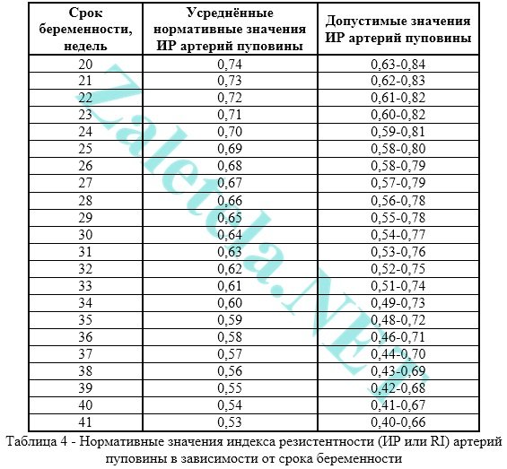
To assess blood flow in the uterine arteries, the resistance index (IR or RI) is calculated.

Often, pregnancy-induced hypertension develops due to impaired uterine blood flow. The expectant mother's body independently increases blood pressure to increase blood flow into the intervillous space. This is how the mother, without realizing it, helps the baby. Thus, it is necessary to improve blood flow and hypertension will disappear on its own.
Impaired blood flow in the uterine arteries is when the value of IR, PI or SDO is greater than normal.
The pulsation index (PI) of the uterine arteries should be within the following limits.

Indicators in the right and left uterine artery may differ slightly from each other. If both indicators are within normal limits, then this picture is not considered a negative phenomenon.
Deviation of blood flow indicators from the norm in two uterine arteries at once indicates a violation of the uteroplacental circulation. This situation requires specific treatment - move more (regularly go swimming or do gymnastics for pregnant women).
Violation of blood flow in only one uterine artery indicates an asymmetry of uteroplacental blood flow. If the pregnancy proceeds normally and the baby develops in accordance with the term, then the placenta is fulfilling its functions.
You should be aware that at 18-21 weeks there may be a temporary disruption of blood flow in the uterine arteries. This phenomenon is explained by the fact that the adaptive physiological process of cytotrophoblast invasion has not yet been completely completed. Therefore, if abnormalities are detected in the uterine arteries, a repeat Doppler ultrasound should be performed after 2-3 weeks, i.e. observe the blood flow over time.
The systole-diastolic ratio (SDR) in the uterine arteries should be:

Umbilical cord arteries (a. umbilicalis). To obtain true results, the study should be carried out only while the baby is at rest, and only when his heart rate is between 120-160 beats per minute. After all, physiologically it is so laid down that when the heart rate increases, the IR in the umbilical cord artery decreases, and vice versa, when the heart rate decreases, the IR increases.
Measuring blood flow in the umbilical cord arteries should be done while the pregnant woman is lying on her back! Assessment of the severity of umbilical cord blood flow disturbance cannot be objective when the expectant mother is positioned “on her left side.”
The umbilical cord should have two arteries and one vein. If there is an anomaly (a single umbilical cord artery), then the fetus may suffer from a lack of oxygen and nutrients, which is why it lags behind in weight and growth. But it happens that the fetus adapts to such an existence and does not experience a deficiency of necessary substances. Such babies are born with low weight, but absolutely viable. Therefore, if there is one umbilical cord artery and the blood flow in it is not impaired, then there is no cause for concern. But if the blood flow in a single artery is impaired, inpatient treatment should be carried out to improve blood flow and, if necessary, early delivery (if the fetus is severely delayed in development).
The most widely used method for assessing the nature of blood flow in the umbilical cord arteries is the resistance index. The readings in both umbilical cord arteries should be almost the same.
Disturbance of blood flow in the umbilical cord is when the value of IR, PI or SDO in the umbilical cord arteries is higher than normal.
The pulsation index (PI or PI) of the umbilical cord arteries must meet the following standards:

Registration of zero and reverse values of diastolic blood flow is pathological. This means that the fetus is in critical condition.
There are only 2-3 days left from the moment permanent reverse values appear until the death of the fetus, so it is necessary to perform a caesarean section as soon as possible in order to save the baby’s life. This is only possible starting from week 28, when the baby is viable.
Systole-diastolic ratio (SDR) in the umbilical cord arteries:

If the blood flow in the umbilical cord is impaired, then, as a rule, fetal development is delayed. If there is no developmental delay now, but the blood flow in the umbilical cord is impaired, then without treatment, the fetus may experience developmental delay.
Middle cerebral artery of the fetus (a. cerebri media). When the fetus suffers, it is observed increase in the values of PI, SDO and speed in the SMA.

Maximum speed (aka V max) average cerebral artery fetus:

Systole-diastolic ratio (SDR) for the middle cerebral artery:

Fetal aorta. It emerges from the left ventricle of the heart, runs along the spine and ends in the lower abdomen, where the aorta divides into two iliac arteries, which provide blood supply to the human legs.
Abnormalities in the blood flow of the aorta can only be detected after 22-24 weeks of pregnancy.
Impaired blood flow is increasing the values of IR, PI and SDO. Critical (indicating fetal death) is considered registration of extremely low values until their complete disappearance.

Changes in the aorta characterize the severity intrauterine hypoxia fetus


Systole-diastolic ratio (SDR) for the fetal aorta:

Ductus venosus (DV). It is studied with enhanced Doppler assessment of blood flow.
During the study, it is necessary not to take into account episodes of hiccup-like respiratory movements of the child and active movement.
Indices are not used to assess the ductus venosus.
Diagnostic criterion pathological condition fetus is considered to be present negative or zero blood flow values during the phase of atrial contraction. Zero or reverse values are recorded for fetal malnutrition, congenital defects of the right heart, and non-immune hydrops fetalis.
Even with critical blood flow in the umbilical cord arteries, but with preserved blood flow in the ductus venosus during the phase of atrial contraction, it is possible to extend gestation until the optimal time for childbirth.
Description of blood flow disorders and their treatment
1st degree
1 A degree– disturbance of blood flow in the uterine arteries, while in the umbilical cord blood flow remains normal.
This degree of blood flow disturbance is not dangerous for the fetus.
Drug treatment for this condition is ineffective. Doctors still prescribe therapy with Actovegin and Curantil. Don't see each other on occasion!
In fact, if blood flow in the uterine arteries is impaired, it is more advisable to simply walk more often in the fresh air (breathing deeply) + eat right + move more (hiking, special exercises for pregnant women, morning exercises, yoga, swimming). And don't sit at the computer for hours! That's all the treatment is.
1 B degree– disturbance of blood flow in the umbilical cord arteries, but hemodynamics in the uterine arteries are normal.
This degree of blood flow impairment requires the use of blood thinning drugs to avoid developmental delay and fetal hypoxia.
In this case, treatment is prescribed aimed at improving blood circulation (the drug Placenta compositum, Curantil or Trental). Actovegin is prescribed as an antihypoxant that improves oxygen supply to the fetus.
A blood test for clotting ability (coagulogram) is also prescribed. In case of increased blood clotting, it is necessary to take stronger blood-thinning drugs than Curantil (for example, heparin or a product containing acetylsalicylic acid).
Degree I of the disorder does not lead to fetal death. Systematic monitoring of the nature of blood flow is carried out (every 2 weeks) “plus” monitoring of fetal CTG (after 28 weeks of pregnancy). In addition, be sure to monitor the blood pressure of a pregnant woman.
2nd degree– simultaneous disturbance of blood flow in the uterine arteries and in the umbilical cord, which does not reach critical values (when blood flow is preserved in the venous duct).
In this condition, it is mandatory to prescribe drug treatment in a hospital, where round-the-clock monitoring of the condition of the fetus is provided. It is also necessary to monitor the state of blood flow by performing Doppler + CTG every 2 days.
In grade II, hemodynamic disturbances are rare, but cases of intrauterine mortality may occur.
3rd degree– critical disturbances of blood flow in the umbilical cord with intact or impaired blood flow in the uterine arteries. A critical violation is understood as registration of reverse diastolic blood flow or its absence at all.
The third degree of violation poses a danger to the child’s health, because in half of the cases the baby’s intrauterine death occurs. Therefore, if a 3rd degree of blood flow disorder is detected, it is necessary to urgently perform a cesarean section in order to save the baby’s life, because at this stage of the disorder, treatment is not effective.
Conservative (natural) childbirth in the 3rd degree can lead to perinatal death of the child.
The cost of Doppler ultrasound in a private clinic is about 1,200 rubles.
A clearly functioning “mother-placenta-baby” system is the key to the health of a woman expecting a new addition to the family and her baby. A failure in this system, resulting in impaired blood flow, can lead to negative consequences for a child, the reversibility of which is often simply impossible. Violation is fraught with delayed development of the fetus in the womb. The consequences of this disorder also include hypoxia, malformations and even embryonic death.
What is Doppler?
An additional circle of blood circulation in a pregnant woman requires additional examination by a specialist. This examination is called Doppler ultrasound. Doppler is an ultrasound diagnosis of the intensity of blood flow in different vessels. Diagnosis is carried out in the third trimester of pregnancy. It is at this time that Doppler shows almost 100% reliable results. In some cases, Doppler measurements are performed at twenty weeks.
By comparing the information received on the device and guided by blood flow standards, the diagnostician determines whether the child is experiencing oxygen starvation or not.
Doppler testing has its own approved standards, which include: index of vascular resistance of the uterus, umbilical cord, aorta and fetal cerebral artery. Independent decoding and comparison of data obtained after diagnosis and Doppler measurements is a thankless task. Only a doctor can calculate the vascular resistance index using the appropriate formula.

What should you do if the doctor, having deciphered the Doppler data and compared it with the norms, notes a violation of the blood flow of the pregnant woman? Well, definitely don’t panic and don’t get nervous. It won't be good for the child either. Timely prescribed treatment effectively combats blood flow diseases.
Severity of blood flow disorders during pregnancy
Disturbances in the circulation of blood through the blood vessels during pregnancy vary in severity.
In the first degree, blood flow disturbance does not reach critical values. The fetal hemodynamics are positive.
The hemodynamics of the fetus in the second degree of the disease is impaired. In half of the cases, the maximum speed of blood movement through all heart valves is reduced. In this case, blood flow is disrupted both in the child and in the arteries of the uterus of the expectant mother. In a very short period, the second degree can develop into the third.
The third degree is destructive for the child. Its diagnosis states the critical state of the blood supply to the fetus. Intracardiac hemodynamics at this stage have profound changes. Fetal hypoxia is most likely in this case.

Symptoms of impaired blood flow
Can a pregnant woman experience blood flow problems? There are certain symptoms. But, for example, in the first stage, placental insufficiency does not manifest itself in any way. It can only be diagnosed by ultrasound. A second degree symptom is a change in the baby’s behavior. He is either too active or, conversely, inactive. Secondary signs of blood flow disease may include protein excretion in the urine, insufficient or excessive amounts of amniotic fluid, swelling, gestosis (late toxicosis), pressure surges, sudden weight gain.
Bloody discharge from the birth canal is the most danger sign disorders associated with placental abruption. In this situation, only ambulance specialists.

Consequences of impaired blood flow during pregnancy
The consequences of impaired blood flow are very sad if treatment is not prescribed in time. This is, at a minimum, acute or chronic hypoxia, as well as intrauterine growth retardation. More severe complications: premature birth; pregnancy fading; miscarriage; development of congenital pathologies, including those incompatible with life; intrauterine fetal death.
To prevent the disastrous consequences of impaired blood flow, we need, first of all, thorough prevention.
In order for the baby to be fully nourished, a woman must consume a balanced diet during pregnancy. These are products with the maximum possible amount of vitamins and microelements. High-quality proteins, carbohydrates and fats. Frequent consumption of water (more than one liter) is also required. Except in cases where the expectant mother is prone to swelling.

Prevention of pathology
Prevention of blood flow disorders involves monitoring weight changes during pregnancy. An increase of more than 10 kg by the end of pregnancy is considered excessive.
If a pregnant woman is at risk (under 17 years of age or over 36 years of age; with bad habits; having chronic diseases etc.), then prevention should include taking medications, preventing blood flow diseases.
A woman who dreams of becoming the mother of a healthy baby in the future should, already during pregnancy planning, analyze her lifestyle and, if possible, eliminate potential risks.
Video on the topic of the article
Blood flow disorders in the uterine arteries, umbilical cord, placenta during pregnancy (NMPC)
Currently, assessment of uteroplacental blood flow is a mandatory study in generally accepted standards for the management of pregnant women.
Thanks to triple ultrasound screening, which is performed on expert-class devices, obstetricians-gynecologists are able to timely recognize the degree of disturbance of uteroplacental blood flow (UPBF), and, most importantly, choose the necessary tactics for managing the patient.
How does the circulatory system between mother and fetus work?
Many people mistakenly think that only the placenta is responsible for blood flow in the mother-fetus system. In fact, this is too superficial a judgment, because everything is much more complicated.

The uteroplacental blood flow system is an anatomically complex complex of the placenta, as well as the blood vessels of the mother and fetus.
- Levels of the uteroplacental system: The main vessels bringing blood to the placental complex are the terminal branches of the uterine arteries.
- The placenta is the central link in the uteroplacental system. It is here that the most complex processes of blood transfer from mother to child occur. Everyone has long known the postulate that maternal and fetal blood do not mix. But how does this happen? All this is achieved thanks to its complex anatomical structure. The placenta is very firmly attached to the inner wall of the uterus with the help of so-called villi. These “outgrowths” of placental tissue are, as it were, immersed in the thickness of the uterine mucosa. Placental villi are embedded in the walls of the uterine vessels and are practically “washed” with maternal blood. It is here, at the cellular level, that complex processes of diffusion of maternal and fetal blood occur, separated from each other by only a few layers of cells. This is called the “blood-placental barrier,” which literally means “the barrier between the mother’s blood and the placenta.” In addition, it is in the placenta that two blood flows “meet”: from mother to child and vice versa. Such a complex and fragile system cannot but arouse admiration!
- The umbilical cord vessels are the third level in the complex system of blood flow between mother and child. The umbilical cord contains three vessels: two arteries and one vein. The hemodynamics (blood circulation) of the fetus is designed in such a way that the arteries bring blood to the baby’s organs and tissues, and the vein, on the contrary, performs the function of returning blood to the placenta. Disruption of blood flow at this level is called “fetal-placental”; it is the most severe option for the fetus in terms of prognosis.
Video: series of lectures on fetal circulation
Reasons that can lead to disruption of blood flow in the mother-placenta-fetus system
- in a pregnant woman. The decrease leads to an acceleration of blood flow in all vessels, including the uterine arteries. This happens for one simple reason: with anemia, the body tries to increase the delivery of oxygen to the tissues by increasing the speed of circulating blood. This is a compensatory reaction. The same thing happens in the uteroplacental system.
- Pathology of placenta attachment (low placentation, presentation) is characterized by reduced blood flow, since in the area of the lower segment of the uterus the muscle layer is much thinner than in other areas. A similar situation develops when the placenta is attached to the area of the uterine scar (usually after the previous caesarean section). The thinned area of the scar cannot fully provide blood flow, so the amount of blood that flows to the fetus may be insufficient for the normal functioning of the developing organism.
- (late toxicosis) is one of the most common causes of disruption of the blood supply to the uteroplacental system, since this obstetric complication causes damage to small vessels.
- Various infectious diseases suffered during pregnancy. Some microorganisms infect the placenta and cause pathological changes in its tissue, which can cause placental insufficiency.


How dangerous is NMPC for the fetus?

– one of the main consequences of NMPK
Reduced blood supply to the fetus can cause the following obstetric complications:
- Reduced weight and size (intrauterine growth retardation syndrome);
- The disorder, in addition to tachycardia (rapid rhythm) and bradycardia (slow rhythm), arrhythmia can also develop as a result of a disturbance in the electrolyte composition of the blood;
- Violation of the acid-base balance in the fetus’s body (change);
- Pathological functioning of the child’s hormonal system;
- A pronounced decrease in fat depots, which is also manifested by low fetal body weight;
- Threat of miscarriage;
- A critical disruption of blood flow can cause intrauterine fetal death.
Degrees of disturbance of uteroplacental circulation
Changes can occur at each level of the system, which is the basis of the classification:
- 1 A - characterized by changes in blood flow in one of the uterine arteries, while other indicators of the system are normal.
- 1 B – blood circulation is impaired at the fetal-placental level (umbilical cord vessels), while the indicators in the uterine arteries are within normal limits.
- 2 – disturbance at the level of the uterine and umbilical vessels.
- 3 – characterized by critical indicators, up to reverse (reverse) blood flow in the arteries.
This classification is extremely convenient for doctors, as it accurately reflects the level at which changes have occurred. In addition, the degree of disturbance of the uteroplacental circulation also affects the patient’s management tactics.
With grade 1 (A and B), the pregnancy can be maintained and treated with conservative methods, grade 2 is borderline, and with grade 3, emergency surgical delivery may be required.
Diagnostic methods
The “gold standard” for blood flow disorders in the mother-placenta-fetus system at the moment in obstetrics is. This method opens up the possibility of recognizing even the most minor changes, as it is highly sensitive and informative.

Doppler is a type of ultrasound examination, which is based on the use of the Doppler effect. The essence of the study is to measure the frequency of ultrasonic waves reflected from moving objects. When studying blood vessels, the moving particles are the formed elements of blood, in particular, red blood cells, as the most numerous cells. Registration of the obtained data and comparison with the norm is called Dopplerography.
What are the benefits of Doppler ultrasound?
- Modern ultrasound machines make it possible to examine the direction, speed of blood flow in the uterine arteries, and even intraplacental blood flow.
- Thanks to color mapping, it is possible to separately examine blood flow in vessels with different directions of blood (arteries and veins). The direction of arterial blood on the device is reflected in red, and venous blood in blue.
- It has been proven that the use of this method during pregnancy does not have any negative influence on fetal development.
- A unique feature of the method is to predict the development of pregnancy. What does this mean? Many studies have shown that disturbances in the blood flow of the uteroplacental system appear a little earlier than any clinical manifestations of fetal suffering (decreased body weight, change in heart rhythm, etc.). This means that with timely diagnosis of fetal blood supply disorders, the doctor has no a large number of time to make the right decision. An example of this is the detection of a change in blood flow, which is called a “dicrotic notch,” in 90% of cases before the development of clinical manifestations of gestosis (edema, increased blood pressure, the appearance of protein in the urine). Doppler sonography is widely used in obstetric practice, as it gives doctors the opportunity not only to promptly recognize blood flow disturbances, but also to even prevent the development of certain pregnancy complications.
Recently, the results of the use of Doppler sonography not only during pregnancy, but also during childbirth are increasingly being published. Conducted studies prove that the effectiveness of contractions can be assessed by measuring the diastolic blood flow velocity in the uterine arteries. Thus, this research method can even predict weakness or impaired coordination of labor at the beginning of the first stage of labor.
Case from practice
An unusual situation that occurred in the maternity ward forced obstetricians and gynecologists to think about the use of Doppler ultrasound during childbirth.

A 25-year-old woman without any concomitant pathology was admitted into labor. home with regular contractions every 3-5 minutes. First birth, urgent.
According to the anamnesis: the pregnancy proceeded without complications, no fetal pathology was detected, all ultrasound protocols with Doppler measurements were within normal limits.
The first stage of labor proceeded physiologically; when the cervix was fully dilated, the woman in labor was transferred to the delivery room.
However, during pushing, seemingly inexplicable phenomena began to occur: the fetal heartbeat was restored during pushing, but in the intervals between pushing it slowed down significantly. Although, as a rule, everything happens the other way around. In this regard, it was decided to perform an ultrasound to study blood flow in the vessels during childbirth. The result of the study surprised everyone: during the intervals between contractions, the fetus pressed the umbilical cord with its hand, as a result of which the blood flow was significantly disrupted. As hypoxia increased in the child’s body, his arms weakened and he let go of the umbilical cord, meanwhile the blood flow was restored. Taking into account the obtained picture, it was decided to speed up the management of the second stage of labor using auxiliary aids. Thus, thanks to Doppler measurements, doctors managed to avoid serious complications.
Secondary diagnostic methods
In addition to Doppler sonography, there are other research methods that indirectly indicate a violation of blood flow:
- Collection of complaints. If blood circulation is impaired, the fetus experiences hypoxia, which is manifested by an increase in motor activity baby. Typically, pregnant women complain of active fetal movement.
- Listening to the heartbeat with a stethoscope can also help in diagnosis. Slowing or accelerating the rhythm beyond the physiological norm may also indicate hypoxia.
- . Registration of CTG for 40-60 minutes may be sufficient to diagnose signs of intrauterine fetal hypoxia.
- Some ultrasonic indicators ( premature aging placenta, biophysical profile study) can document the unfavorable condition of the fetus.
Important! A slight decrease in estimated fetal weight on ultrasound does not necessarily indicate developmental delay and blood flow disturbances. An isolated measurement of fetal weight is not very informative, since this indicator is also largely influenced by genetic factors. Therefore, it is necessary to take into account the anthropometric indicators of the parents (height, weight) and their birth weight. Only a combination of decreased fetal weight and impaired blood flow during Doppler ultrasound provides grounds for a diagnosis of “intrauterine growth retardation syndrome.”
Treatment of uteroplacental blood flow disorders

In order to improve the blood supply to the fetus, it is necessary to influence several links of pathogenesis at once:
- Improving microcirculation. The following drugs have this property: “Pentoxifylline”, “Actovegin”.
- Maintaining normal blood flow in the vessels and eliminating low blood pressure is achieved through the use of drugs based on hydroxyethyl starch: “Stabizol”, “Infukol”, “Voluven”, “ReoHES”, “Venofundin”. These drugs are an osmotic solution of starches that are capable of retaining fluid in the lumen of blood vessels. Their administration is especially effective for gestosis, when it is necessary to transfer fluid from the intercellular space into the lumen of the vessel. In this way, two goals are achieved at once: normalizing blood flow and reducing the severity of edema.
- Vasodilators help eliminate spasm of arteries and arterioles (smaller vessels). Particularly effective use “Eufillina”, “No-shpy”, “Magnesia” in the form of injections.
- Reducing the tone of the uterus also helps eliminate vasospasm, hypoxia, and also preserve pregnancy if there is a threat premature birth. In this situation, it is prescribed “Magnesia”, “Ginipral”, “Magne B6”.
- Drugs with antioxidant effects help cope with the undesirable effects of hypoxia. Usually prescribed “Tocopherol”, “Ascorbic acid”, “Hophytol”.
- Useful intravenous administration 5% glucose solution, which has detoxifying properties.
- The drug has a protective effect on the placenta “Essentiale”, which saturates the placental tissue with phospholipids. The use of this medicine is especially useful for placental insufficiency.
- When blood flow is disrupted, it is very effective to prescribe drugs that lead to processes of activation of cellular respiration, for example "Cocarboxylase".
- When uterine fibroids and blood flow disturbances are combined, the drug showed good results “Courantil”, it is able to improve blood circulation in the vessels and also prevents the formation of microthrombi. Many studies have shown that taking “Curantil” for large fibroids prevents the development of secondary, inflammatory changes in myomatous nodes, due to improved blood flow in them and the uteroplacental system.
Conclusion
Study of uteroplacental blood flow using Doppler ultrasound is a mandatory study during pregnancy. Practice shows that the use of this method has had a positive effect on the prevention and treatment of many obstetric complications. However, we should not forget that in order to obtain reliable and informative results, blood flow studies must be carried out using expert-class devices by highly qualified specialists.
Video: about fetal hypoxia – a consequence of NMPK
Impaired blood flow during pregnancy is determined using Doppler ultrasound. The examination shows the movement of blood cells through the vessels from the mother through the placenta to the fetus. The further gestational period depends on these parameters; the doctor receives information about the lack of oxygen or nutrients, which is so important for the normal formation of the baby.
Blood flow during pregnancy
The circulatory system between mother and fetus is quite complex, so in medical practice it is customary to divide it into levels to understand the degree of damage. The main compartment of the entire blood flow system is the placental membrane, which supplies the embryo with all the necessary substances. At the same time, there is a hematoplacental barrier, determined by the presence of special cells that do not allow various harmful cellular structures to pass through.The next part of the uterine blood flow is the spiral arteries, which lack a muscular layer and in case of significant blood loss will not be able to contract properly. And the last structure of blood circulation between a woman and an embryo is called the vessels in the umbilical cord. There are only 2 arteries and 1 vein connecting the placenta to the fetus. Disruption of blood flow during pregnancy in this fetal-placental circle causes the most severe consequences.
Causes of blood flow disorders during pregnancy
There are no reliable reasons that lead to disruption of blood flow in the uterine artery. The only known factors predisposing to disruptions in blood circulation between intrauterine development and the mother are:- severe diseases of the endocrine system, diabetes or abnormalities in the functioning of the adrenal glands and thyroid gland;
- poor functioning of the kidneys and lungs;
- cardiovascular pathologies and hypertension;
- anemia caused by a lack of iron, vitamins and anemia;
- increased thrombus formation;
- Infectious diseases can cause inflammation and swelling of blood vessels, which threatens miscarriage at the beginning of pregnancy.
Degrees of blood flow disturbances during pregnancy
Analysis of blood flow during pregnancy, as well as Doppler ultrasound, help determine several degrees of circulatory abnormalities in all vessels between the woman and the baby. In this case, doctors pay attention to infectious lesions, abnormalities in the composition of the amniotic fluid, the condition of the placenta and other factors that influence the diagnosis.Blood flow disturbances occur:
1st degree - the uteroplacental circle functions with minimal deviation, which gives doctors time to use drug therapy and restore circulatory function. In this case, the 1st degree has two subtypes:
- the functioning of the blood flow between the fetus and the placenta is stable, and the uteroplacental circle is malfunctioning, which characterizes degree 1a of disorders;
- Degree 1b disorders are characterized by the opposite situation, when a lack of functioning is observed in the fetal-placental circle.
Grade 3 is characterized by critical marks during diagnostic measures. If poor blood flow is associated with the placenta, then this will be called placental insufficiency, and may be caused by premature detachment or early aging of the shell.
Thus, by classifying disorders, doctors can see a complete diagnostic picture of the blood flow condition and prescribe appropriate treatment.
Normal blood flow during pregnancy - table
To assess the movement of blood in various vessels, several indicators are used:- The RI or resistive index is the difference between blood flow velocities over the entire study period;
- PI or pulsation index - comparison of velocities during one circulatory cycle;
- SDO or systole-distal ratio involves assessing the speed between heart contractions and during the “rest” period.
| Vessel | Index | 28 weeks | 32 weeks | 36 weeks | 40 weeks |
|---|---|---|---|---|---|
| Umbilical artery | FROM TO | 3,1-3,7 | 2,8-3,4 | 2,4-3,0 | 2,2-2,5 |
| IR | 0,65-0,73 | 0,60-0,67 | 0,55-0,63 | 0,51-0,59 | |
| Pi | 1,08-1,09 | 0,95-0,96 | 0,83-0,84 | 0,73-0,74 | |
| Fetal aorta | FROM TO | 6,0-7,6 | 5,7-7,3 | 5,4-7,1 | 5,2-6,8 |
| IR | 0,82-0,88 | 0,80-0,86 | 0,77-0,83 | 0,75-0,81 | |
| Pi | 1,79-2,24 | 1,76-2,20 | 1,74-2,17 | 1,72-2,13 | |
| Internal carotid artery | FROM TO | 5,6-6,5 | 4,7-5,6 | 4,0-4,8 | 3,3-4,1 |
| IR | 0,78-0,88 | 0,74-0,84 | 0,71-0,81 | 0,69-0,78 | |
| Pi | 1,98-2,39 | 1,7-2,06 | 1,44-1,77 | 1,22-1,51 | |
| Uterine artery | FROM TO | 1,7-1,9 | 1,7-1,9 | 1,7-1,9 | 1,7-1,9 |
| IR | 0,46-0,55 | 0,69-0,86 | 0,69-0,86 | 0,69-0,86 | |
| Pi | 0,69-0,86 | 0,69-0,86 | 0,69-0,86 | 0,69-0,86 |
If the norm is exceeded in the arteries of the uterus, this indicates an insufficient supply of oxygen to the baby; in the umbilical cord, this indicates the presence of gestosis and vascular insufficiency. If abnormalities in the aorta are observed, a diagnosis of an abnormal state of intrauterine development is made; additional tests will be required to normalize the vital signs of the fetus.
To improve blood flow, a drug is used that relaxes the muscles of the uterus on an individual basis; Ginipral, Magne-B6 or Eufillin are most often prescribed. Additionally, Verapamil or Isoptin is used to reduce tachycardia.
To normalize blood flow, it is also necessary to balance the woman’s diet, food and, in general, the daily menu should contain the maximum amount of vitamins and nutrients. It is necessary to reduce the emotional and physical stress of the body so that blood circulation parameters stabilize.